

Brief introduction to Positron Emission Tomography
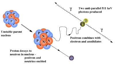
Positron Emission Tomography (PET) is a non-invasive medical imaging technique. A positron
emitter is used to mark a tracer which is injected into a living organism. By e+e- annihilation inside the organism two 511 keV
photons are produced (see figure 1).
 They are detected in coincidence and their line of response is identified (see figure 2).
The reconstruction of several lines of response allows
to retrieve a functional image of the tissue in which the tracer is absorbed.
They are detected in coincidence and their line of response is identified (see figure 2).
The reconstruction of several lines of response allows
to retrieve a functional image of the tissue in which the tracer is absorbed.
 The energy and the time resolution of the used gamma-detectors are two important parameters which determine
the overall performance of the PET scanner. A good energy resolution is crucial in order to
reject photons which have undergone Compton scattering inside the organism and thus changed
their direction. The time resolution of the gamma-detectors determines the minimal width of
the coincidence window and thus the amount of background arising from random coincidences.
Furthermore, the time information can be used to directly improve the image quality especially
for heavier patients using time-of-flight techniques (see figure 3).
The energy and the time resolution of the used gamma-detectors are two important parameters which determine
the overall performance of the PET scanner. A good energy resolution is crucial in order to
reject photons which have undergone Compton scattering inside the organism and thus changed
their direction. The time resolution of the gamma-detectors determines the minimal width of
the coincidence window and thus the amount of background arising from random coincidences.
Furthermore, the time information can be used to directly improve the image quality especially
for heavier patients using time-of-flight techniques (see figure 3).

Figure 1: A positron emitted by the tracer atom annihilates with an electron in the tissue.
Figure 2: Two 511 keV photons which are detected in coincidence form a line of response (LOR).
Figure 3: The TOF-PET principle.
Apart from the requirements on the energy and time resolution a small pixel size of the PET camera, i.e. a high granularity, is required since this mainly determines the spatial resolution.
Back